JavaScript is disabled for your browser. Some features of this site may not work without it.
Buscar en RiuNet
Listar
Mi cuenta
Estadísticas
Ayuda RiuNet
Admin. UPV
Automatic Segmentation of the Retinal Nerve Fiber Layer by Means of Mathematical Morphology and Deformable Models in 2D Optical Coherence Tomography Imaging
Mostrar el registro sencillo del ítem
Ficheros en el ítem
| dc.contributor.author | Berenguer-Vidal, Rafael
|
es_ES |
| dc.contributor.author | Verdú-Monedero, Rafael
|
es_ES |
| dc.contributor.author | Morales-Sánchez, Juan
|
es_ES |
| dc.contributor.author | Sellés-Navarro, Inmaculada
|
es_ES |
| dc.contributor.author | del Amor, Rocío
|
es_ES |
| dc.contributor.author | García-Pardo, José Gabriel
|
es_ES |
| dc.contributor.author | Naranjo Ornedo, Valeriana
|
es_ES |
| dc.date.accessioned | 2022-04-28T18:04:49Z | |
| dc.date.available | 2022-04-28T18:04:49Z | |
| dc.date.issued | 2021-12 | es_ES |
| dc.identifier.uri | http://hdl.handle.net/10251/182267 | |
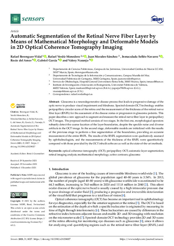
| dc.description.abstract | [EN] Glaucoma is a neurodegenerative disease process that leads to progressive damage of the optic nerve to produce visual impairment and blindness. Spectral-domain OCT technology enables peripapillary circular scans of the retina and the measurement of the thickness of the retinal nerve fiber layer (RNFL) for the assessment of the disease status or progression in glaucoma patients. This paper describes a new approach to segment and measure the retinal nerve fiber layer in peripapillary OCT images. The proposed method consists of two stages. In the first one, morphological operators robustly detect the coarse location of the layer boundaries, despite the speckle noise and diverse artifacts in the OCT image. In the second stage, deformable models are initialized with the results of the previous stage to perform a fine segmentation of the boundaries, providing an accurate measurement of the entire RNFL. The results of the RNFL segmentation were qualitatively assessed by ophthalmologists, and the measurements of the thickness of the RNFL were quantitatively compared with those provided by the OCT inbuilt software as well as the state-of-the-art methods. | es_ES |
| dc.description.sponsorship | This work was partially funded by Spanish National projects AES2017-PI17/00771 and AES2017-PI17/00821 (Instituto de Salud Carlos III), PID2019-105142RB-C21 (AI4SKIN) (Spanish Ministry of Economy and Competitiveness), PTA2017-14610-I (State Research Spanish Agency), regional project 20901/PI/18 (Fundacion Seneca) and Polytechnic University of Valencia (PAID-01-20). | es_ES |
| dc.language | Inglés | es_ES |
| dc.publisher | MDPI AG | es_ES |
| dc.relation.ispartof | Sensors | es_ES |
| dc.rights | Reconocimiento (by) | es_ES |
| dc.subject | Optical coherence tomography (OCT) | es_ES |
| dc.subject | Peripapillary OCT | es_ES |
| dc.subject | Automatic layer segmentation | es_ES |
| dc.subject | Retinal imaging analysis | es_ES |
| dc.subject | Mathematical morphology | es_ES |
| dc.subject | Active contours | es_ES |
| dc.subject | Glaucoma | es_ES |
| dc.subject.classification | TEORIA DE LA SEÑAL Y COMUNICACIONES | es_ES |
| dc.title | Automatic Segmentation of the Retinal Nerve Fiber Layer by Means of Mathematical Morphology and Deformable Models in 2D Optical Coherence Tomography Imaging | es_ES |
| dc.type | Artículo | es_ES |
| dc.identifier.doi | 10.3390/s21238027 | es_ES |
| dc.relation.projectID | info:eu-repo/grantAgreement/AEI/Plan Estatal de Investigación Científica y Técnica y de Innovación 2017-2020/PID2019-105142RB-C21/ES/CARACTERIZACION DE NEOPLASIAS DE CELULAS FUSIFORMES EN IMAGENES HISTOLOGICAS/ | es_ES |
| dc.relation.projectID | info:eu-repo/grantAgreement/MINECO//AES2017-PI17%2F00771/ | es_ES |
| dc.relation.projectID | info:eu-repo/grantAgreement/EC/H2020/732613/EU | es_ES |
| dc.relation.projectID | info:eu-repo/grantAgreement/MINECO//AES2017-PI17%2F00821/ | es_ES |
| dc.relation.projectID | info:eu-repo/grantAgreement/f SéNeCa//20901%2FPI%2F18/ | es_ES |
| dc.relation.projectID | info:eu-repo/grantAgreement/UPV//PAID-01-20 nº21589//Programa de Apoyo para la Investigación y Desarrollo de la Universitat Politècnica de València/ | es_ES |
| dc.relation.projectID | info:eu-repo/grantAgreement/AEI//PTA2017-14610-I//AYUDA TECNICO DE APOYO MINISTERIO-GARCIA PARDO/ | es_ES |
| dc.rights.accessRights | Abierto | es_ES |
| dc.contributor.affiliation | Universitat Politècnica de València. Departamento de Comunicaciones - Departament de Comunicacions | es_ES |
| dc.description.bibliographicCitation | Berenguer-Vidal, R.; Verdú-Monedero, R.; Morales-Sánchez, J.; Sellés-Navarro, I.; Del Amor, R.; García-Pardo, JG.; Naranjo Ornedo, V. (2021). Automatic Segmentation of the Retinal Nerve Fiber Layer by Means of Mathematical Morphology and Deformable Models in 2D Optical Coherence Tomography Imaging. Sensors. 21(23):1-30. https://doi.org/10.3390/s21238027 | es_ES |
| dc.description.accrualMethod | S | es_ES |
| dc.relation.publisherversion | https://doi.org/10.3390/s21238027 | es_ES |
| dc.description.upvformatpinicio | 1 | es_ES |
| dc.description.upvformatpfin | 30 | es_ES |
| dc.type.version | info:eu-repo/semantics/publishedVersion | es_ES |
| dc.description.volume | 21 | es_ES |
| dc.description.issue | 23 | es_ES |
| dc.identifier.eissn | 1424-8220 | es_ES |
| dc.identifier.pmid | 34884031 | es_ES |
| dc.identifier.pmcid | PMC8659929 | es_ES |
| dc.relation.pasarela | S\451591 | es_ES |
| dc.contributor.funder | Agencia Estatal de Investigación | es_ES |
| dc.contributor.funder | COMISION DE LAS COMUNIDADES EUROPEA | es_ES |
| dc.contributor.funder | Universitat Politècnica de València | es_ES |
| dc.contributor.funder | Ministerio de Economía y Competitividad | es_ES |
| dc.contributor.funder | Fundación Séneca-Agencia de Ciencia y Tecnología de la Región de Murcia | es_ES |


