JavaScript is disabled for your browser. Some features of this site may not work without it.
Buscar en RiuNet
Listar
Mi cuenta
Estadísticas
Ayuda RiuNet
Admin. UPV
Atrial low voltage areas: A comparison between atrial fibrillation and sinus rhythm
Mostrar el registro sencillo del ítem
Ficheros en el ítem
| dc.contributor.author | Andrés Lahuerta, Ana
|
es_ES |
| dc.contributor.author | Roberto, Carlos
|
es_ES |
| dc.contributor.author | Saiz Rodríguez, Francisco Javier
|
es_ES |
| dc.contributor.author | Cano, Óscar
|
es_ES |
| dc.contributor.author | Martínez-Mateu, Laura
|
es_ES |
| dc.contributor.author | Alonso, Pau
|
es_ES |
| dc.contributor.author | Saurí, Assumpció
|
es_ES |
| dc.contributor.author | Quesada, Aurelio
|
es_ES |
| dc.contributor.author | Osca, Joaquín
|
es_ES |
| dc.date.accessioned | 2022-05-05T18:03:44Z | |
| dc.date.available | 2022-05-05T18:03:44Z | |
| dc.date.issued | 2022 | es_ES |
| dc.identifier.issn | 1897-5593 | es_ES |
| dc.identifier.uri | http://hdl.handle.net/10251/182394 | |
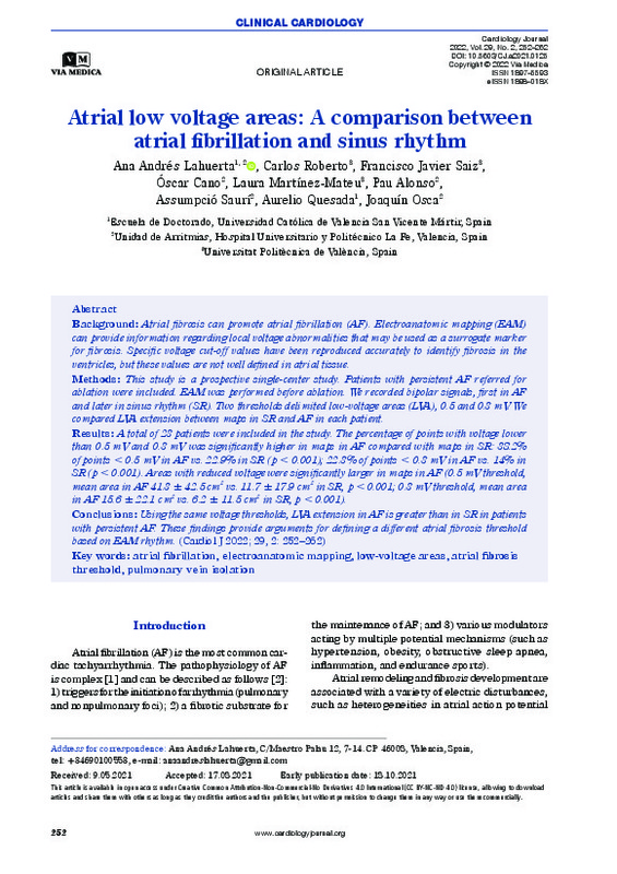
| dc.description.abstract | [EN] Background: Atrial fibrosis can promote atrial fibrillation (AF). Electroanatomic mapping (EAM) can provide information regarding local voltage abnormalities that may be used as a surrogate marker for fibrosis. Specific voltage cut-off values have been reproduced accurately to identify fibrosis in the ventricles, but these values are not well defined in atrial tissue. Methods: This study is a prospective single-center study. Patients with persistent AF referred for ablation were included. EAM was performed before ablation. We recorded bipolar signals, first in AF and later in sinus rhythm (SR). Two thresholds delimited low-voltage areas (LVA), 0.5 and 0.3 mV. We compared LVA extension between maps in SR and AF in each patient. Results: A total of 23 patients were included in the study. The percentage of points with voltage lower than 0.5 mV and 0.3 mV was significantly higher in maps in AF compared with maps in SR: 38.2% of points < 0.5 mV in AF vs. 22.9% in SR (p < 0.001); 22.3% of points < 0.3 mV in AF vs. 14% in SR (p < 0.001). Areas with reduced voltage were significantly larger in maps in AF (0.5 mV threshold, mean area in AF 41.3 ± 42.5 cm2 vs. 11.7 ± 17.9 cm2 in SR, p < 0.001; 0.3 mV threshold, mean area in AF 15.6 ± 22.1 cm2 vs. 6.2 ± 11.5 cm2 in SR, p < 0.001). Conclusions: Using the same voltage thresholds, LVA extension in AF is greater than in SR in patients with persistent AF. These findings provide arguments for defining a different atrial fibrosis threshold based on EAM rhythm. | es_ES |
| dc.language | Inglés | es_ES |
| dc.publisher | VM Media Sp zo.o. - VMGroup SK | es_ES |
| dc.relation.ispartof | Cardiology Journal | es_ES |
| dc.rights | Reconocimiento - No comercial - Sin obra derivada (by-nc-nd) | es_ES |
| dc.subject | Atrial fibrillation | es_ES |
| dc.subject | Electroanatomic mapping | es_ES |
| dc.subject | Low-voltage areas | es_ES |
| dc.subject | Atrial fibrosis threshold | es_ES |
| dc.subject | Pulmonary vein isolation | es_ES |
| dc.subject.classification | TECNOLOGIA ELECTRONICA | es_ES |
| dc.title | Atrial low voltage areas: A comparison between atrial fibrillation and sinus rhythm | es_ES |
| dc.type | Artículo | es_ES |
| dc.identifier.doi | 10.5603/CJ.a2021.0125 | es_ES |
| dc.rights.accessRights | Abierto | es_ES |
| dc.contributor.affiliation | Universitat Politècnica de València. Departamento de Ingeniería Electrónica - Departament d'Enginyeria Electrònica | es_ES |
| dc.description.bibliographicCitation | Andrés Lahuerta, A.; Roberto, C.; Saiz Rodríguez, FJ.; Cano, Ó.; Martínez-Mateu, L.; Alonso, P.; Saurí, A.... (2022). Atrial low voltage areas: A comparison between atrial fibrillation and sinus rhythm. Cardiology Journal. 29(2):252-262. https://doi.org/10.5603/CJ.a2021.0125 | es_ES |
| dc.description.accrualMethod | S | es_ES |
| dc.relation.publisherversion | https://doi.org/10.5603/CJ.a2021.0125 | es_ES |
| dc.description.upvformatpinicio | 252 | es_ES |
| dc.description.upvformatpfin | 262 | es_ES |
| dc.type.version | info:eu-repo/semantics/publishedVersion | es_ES |
| dc.description.volume | 29 | es_ES |
| dc.description.issue | 2 | es_ES |
| dc.identifier.pmid | 34642920 | es_ES |
| dc.identifier.pmcid | PMC9007488 | es_ES |
| dc.relation.pasarela | S\462083 | es_ES |
| dc.subject.ods | 03.- Garantizar una vida saludable y promover el bienestar para todos y todas en todas las edades | es_ES |


