JavaScript is disabled for your browser. Some features of this site may not work without it.
Buscar en RiuNet
Listar
Mi cuenta
Estadísticas
Ayuda RiuNet
Admin. UPV
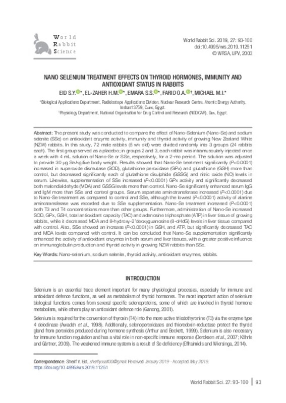
Nano selenium treatment effects on thyroid hormones, immunity and antioxidant status in rabbits
Mostrar el registro sencillo del ítem
Ficheros en el ítem
| dc.contributor.author | Eid, Sherif Yousif
|
es_ES |
| dc.contributor.author | El-Zaher, Hussein Mustafa
|
es_ES |
| dc.contributor.author | Emara, Sana Sayed
|
es_ES |
| dc.contributor.author | Farid, Omar Abdel-Hamed
|
es_ES |
| dc.contributor.author | Michael, Michael Ibrahim
|
es_ES |
| dc.date.accessioned | 2019-10-02T12:09:32Z | |
| dc.date.available | 2019-10-02T12:09:32Z | |
| dc.date.issued | 2019-06-28 | |
| dc.identifier.issn | 1257-5011 | |
| dc.identifier.uri | http://hdl.handle.net/10251/127019 | |
| dc.description.abstract | [EN] The present study was conducted to compare the effect of Nano-Selenium (Nano-Se) and sodium selenite (SSe) on antioxidant enzyme activity, immunity and thyroid activity of growing New Zealand White (NZW) rabbits. In this study, 72 male rabbits (5 wk old) were divided randomly into 3 groups (24 rabbits each). The first group served as a placebo; in groups 2 and 3, each rabbit was intramuscularly injected once a week with 4 mL solution of Nano-Se or SSe, respectively, for a 2-mo period. The solution was adjusted to provide 30 μg Se/kg/live body weight. Results showed that Nano-Se treatment significantly (P<0.0001) increased in superoxide dismutase (SOD), glutathione peroxidase (GPx) and glutathione (GSH) more than control, but decreased significantly each of glutathione disulphide (GSSG) and nitric oxide (NO) levels in serum. Likewise, supplementation of SSe increased (P<0.0001) GPx activity and significantly decreased both malondialdehyde (MDA) and GSSG levels more than control. Nano-Se significantly enhanced serum IgG and IgM more than SSe and control groups. Serum aspartate aminotransferase increased (P<0.0001) due to Nano-Se treatment as compared to control and SSe, although the lowest (P<0.0001) activity of alanine aminotransferase was recorded due to SSe supplementation. Nano-Se treatment increased (P<0.0001) both T3 and T4 concentrations more than other groups. Furthermore, administration of Nano-Se increased SOD, GPx, GSH, total antioxidant capacity (TAC) and adenosine triphosphate (ATP) in liver tissue of growing rabbits, while it decreased MDA and 8-hydroxy-2’deoxyguanosine (8-oHdG) levels in liver tissue compared with control. Also, SSe showed an increase (P<0.0001) in GSH, and ATP, but significantly decreased TAC and MDA levels compared with control. It can be concluded that Nano-Se supplementation significantly enhanced the activity of antioxidant enzymes in both serum and liver tissues, with a greater positive influence on immunoglobulin production and thyroid activity in growing NZW rabbits than SSe. | es_ES |
| dc.language | Inglés | es_ES |
| dc.publisher | Universitat Politècnica de València | |
| dc.relation.ispartof | World Rabbit Science | |
| dc.rights | Reserva de todos los derechos | es_ES |
| dc.subject | Nano-selenium | es_ES |
| dc.subject | Sodium selenite | es_ES |
| dc.subject | Thyroid activity | es_ES |
| dc.subject | Antioxidant enzymes | es_ES |
| dc.subject | Rabbits | es_ES |
| dc.title | Nano selenium treatment effects on thyroid hormones, immunity and antioxidant status in rabbits | es_ES |
| dc.type | Artículo | es_ES |
| dc.date.updated | 2019-10-02T11:49:37Z | |
| dc.identifier.doi | 10.4995/wrs.2019.11251 | |
| dc.rights.accessRights | Abierto | es_ES |
| dc.description.bibliographicCitation | Eid, SY.; El-Zaher, HM.; Emara, SS.; Farid, OA.; Michael, MI. (2019). Nano selenium treatment effects on thyroid hormones, immunity and antioxidant status in rabbits. World Rabbit Science. 27(2):93-100. https://doi.org/10.4995/wrs.2019.11251 | es_ES |
| dc.description.accrualMethod | SWORD | es_ES |
| dc.relation.publisherversion | https://doi.org/10.4995/wrs.2019.11251 | es_ES |
| dc.description.upvformatpinicio | 93 | es_ES |
| dc.description.upvformatpfin | 100 | es_ES |
| dc.type.version | info:eu-repo/semantics/publishedVersion | es_ES |
| dc.description.volume | 27 | |
| dc.description.issue | 2 | |
| dc.identifier.eissn | 1989-8886 | |
| dc.description.references | Abd-Allah S., Hashem K.S. 2015. Selenium nanoparticles increase the testicular antioxidant activity and spermatogenesis in male rats as compared to ordinary selenium. Int. J. Adv. Res., 3: 792-802. https://doi.org/10.22038/ijbms.2018.26023.6397 | es_ES |
| dc.description.references | Abu-Qare A., Abou-Donia, M. 2000. Increased 8-hydroxy-2'- deoxyguanosine, a biomarker of oxidative DNA damage in rat urine following a single dermal dose of DEET (N, N-diethylm-toluamide), and permethrin, alone and in combination. Toxicol. Lett., 117: 151-60. https://doi.org/10.1016/S0378-4274(00)00257-5 | es_ES |
| dc.description.references | AOAC. 2000. Official Methods of Analysis of AOAC International, Gaithersburg MA, USA. Association of Official Analytical chemist. | es_ES |
| dc.description.references | Aurthor J.R., Beckett G.J. 1999. Thyroid function. Br. Med. Bull. 55: 658-668. | es_ES |
| dc.description.references | Awadeh F.T., Kincaid J.R., Johnson K.A. 1998. Effect of level and source of dietary selenium on concentrations of thyroid hormones and immunoglobulins in beef cows and calves. J. Anim. Sci. 76: 1204-1215. | es_ES |
| dc.description.references | Bickhardt K., Ganterm M., Sallmann P. 1999. Investigation of the manifestation of vitamin E and selenium deficiency in sheep and goats. Dtsch. Tierarztl. Wochenschr., 106: 242-247. | es_ES |
| dc.description.references | Changguang L., Jingui Z., Jinyu L. 2013. Effects of different sources and supplemental dosages of selenium on growth performance, immunity and thyroid hormones. China Feed, 21: 007 | es_ES |
| dc.description.references | Dercksen D.P., Counotte G.H., Hazebroek M.K., Arts W., Van R.T. 2007. Selenium requirements of dairy goats. Tijdschr. Diergeneeskd, 132: 468-471. | es_ES |
| dc.description.references | Effraimidis G., Wiersinga M. 2014. Mechanisms in endocrinology: Autoimmune thyroid disease: Old and new players. Eur. J. Endocrinol., 170: 241-252. | es_ES |
| dc.description.references | Everett S.A., Dennis M.F., Tozer G.M., Prise V.E., Wardman P., Stratford M.R. 1995. Nitric oxide in biological fluids: analysis of nitrite and nitrate by high-performance ion chromatography. J. Chromatogr. A, 706: 37-42. https://doi.org/10.1016/0021-9673(95)00078-2. | es_ES |
| dc.description.references | Ganong W.F. 2001. The thyroid gland: In: Review of Medical Physiology. New York: Lange Medical Books/McGraw-Hill, 307-321. | es_ES |
| dc.description.references | Goldberg D.M., Spoone R.J. 1983. Assay of glutathione reductase In: Bergmeyen H.V. (ed). Methods of enzymatic analysis. Florida: Verlag Chemie Deerfield Beach, 258-265. | es_ES |
| dc.description.references | Habig W., Pabst M., Jakoby W. 1974. Glutathione S-transferases: the first enzymatic step in mercapturic acid formation. J. Biol. Chem. 249: 7130-7139. | es_ES |
| dc.description.references | Hao X., Ling Q., Hong F. 2014. Effects of dietary selenium on the pathological changes and oxidative stress in loach (Paramisgurnus dabryanus). Fish Physiol. Biochem., 40: 1313-1323. https://doi.org/10.1007/s10695-014-9926-7. | es_ES |
| dc.description.references | Holmgren A., Johansson C., Berndt C., Lonn M.E., Hudemann C., Lillig C.H. 2005. Thiol redox control via thioredoxin and glutaredoxin systems. Biochem. Soc. Trans., 33: 1375-1377. | es_ES |
| dc.description.references | Huang B., Zhang J., Hou J., Chen C. 2003. Free radical scavenging efficiency of nano-Se in vitro. Free Radic. Biol. Med., 35: 805-813. https://doi.org/10.1016/S0891-5849(03)00428-3 | es_ES |
| dc.description.references | Jayatilleke E., Shaw S. 1993. A high-performance liquid chromatographic assay for reduced and oxidized glutathione in biological samples. Anal. Biochem., 214: 452-457. | es_ES |
| dc.description.references | Jia X., Li N., Chen J. 2005. A subchronic toxicity study of elemental Nano-Se in Sprague-Dawley rats. Life Sci., 76: 1989-2003. https://doi.org/10.1016/j.lfs.2004.09.026 | es_ES |
| dc.description.references | Kasai H. 2002. Chemistry-based studies on oxidative DNA damage: formation, repair, and mutagenesis. Free Radic. Biol. Med., 33: 450-456. | es_ES |
| dc.description.references | Karatepe M. 2004. Simultaneous determination of ascorbic acid and free malondialdehyde in human serum by HPLC-UV. Chromatographic Line, 12: 362-365. | es_ES |
| dc.description.references | Köhrle J., Gartner R. 2009. Selenium and thyroid. Best. Pract. Res. Clin. En. 23: 815-827. https://doi.org/10.1016/j.beem.2009.08.002 | es_ES |
| dc.description.references | Köhrle J., Jakob F., Contempré B., Dumont J.E. 2007. Selenium, the thyroid, and the endocrine system. Endocrinol. Rev., 26: 944-984. https://doi.org/10.1210/er.2001-0034 | es_ES |
| dc.description.references | Koracevic D., Koracevic G., Djordjevic V., Andrejevic S., Cosic V. 2001. Method for the measurement of antioxidant activity in human fluids. J. Clin. Pathol., 54: 356-361. | es_ES |
| dc.description.references | Kumar N., Garg A.K., Mudgal V. 2008. Effect of different levels of selenium supplementation on growth rate, nutrient utilization, blood metabolic profile, and immune response in lambs. Biol. Trace Elem. Res., 126: 44-56. | es_ES |
| dc.description.references | Lawrence R.A., Burk R.F. 1976. Glutathione peroxidase activity in selenium deficient rat liver. Biochem. Biophys. Res. Commun.,71: 952-958. | es_ES |
| dc.description.references | Liao C.D, Hung WL, Jan K.C, Yeh A.I, Ho C.T, Hwang L.S. 2010. Nano/sub-microsized lignan glycosides from sesame meal exhibit higher transport and absorption efficiency in Caco-2 cell monolayer. Food Chem., 119: 896-902. http://doi.org/10.1016/j.foodchem.2009.07.056 | es_ES |
| dc.description.references | Liu H.Y., Jiang Y., Luo W. 2006. A simple and rapid determination of ATP, ADP and AMP concentrations in pericarp tissue of litchi fruit by high performance liquid chromatography. Food Technol. Biotech., 44: 531-534. | es_ES |
| dc.description.references | Lodovici M., Casalini C., Briani C., Dolara P. 1997. Oxidative liver DNA damage in rats treated with pesticide mixtures. Toxicology, 117: 55-60. | es_ES |
| dc.description.references | Marin-Guzman J, Mahan D.C, Pate J.L. 2000. Effect of dietary selenium and vitamin E on spermatogenic development in boars. J. Anim .Sci., 78: 1536-1543. | es_ES |
| dc.description.references | Mohapatra P., Swain R. K., Mishra S. K., Behera T., Swain P., Mishra S. S., Behura N.C., Sabat S.C., Sethy K., Dhama K., Jayasankar P. 2014. Effects of dietary nano-selenium on tissue selenium deposition, antioxidant status and immune functions in layer chicks. Int. J. Pharmacol., 10: 160-167. https://doi.org/10.3923/ijp.2014.160.167 | es_ES |
| dc.description.references | Montgomery J.B., Wichtel J.J., Wichtel M.G., McNiven M.A., McClure J.T., Markham F., Horohov D.W. 2012. Effects of selenium source on measures of selenium status and immune function in horses. Canad. J. Vet. Res., 76: 281-291. | es_ES |
| dc.description.references | Nasirpour M., Sadeghi A.A., Chamani M. 2017. Effects of nano-selenium on the liver antioxidant enzyme activity and immunoglobulins in male rats exposed to oxidative stress. J. Livestock Sci., 8: 81-87. | es_ES |
| dc.description.references | NRC. 1977. Nutrient requirements of rabbits. National Research Council, Natl. Acad. Press, Washington, DC, USA. | es_ES |
| dc.description.references | Paglia D.E., Valentine W.N. 1967. Studies on the quantitative and qualitative characterization of erythrocyte glutathione peroxidase. J. Lab. Clin. Med, 70: 158-169. | es_ES |
| dc.description.references | Papadoyannis L.N., Samanidou V.F., Nitsos C.C. 1999. Simultaneous determination of nitrite and nitrate in drinking water and human serum by high-performance anionexchange chromatography and UV detection. J. Liq. Chrom. Rel. Techno., 22: 2023-2041. | es_ES |
| dc.description.references | Qin S., Chen F., Guo Y., Huang B., Zhang J., Ma J. 2014. Effects of nano-selenium on kidney selenium contents, glutathione peroxidase activities and GPx-1 mRNA expression in mice. Adv. Mater. Res., 1051: 383-387. https://doi.org/10.4028/www.scientific.net/AMR.1051.383 | es_ES |
| dc.description.references | Qin F., Chen F., Zhao F., Jin T., Ma J. 2016. Effects of Nanoselenium on Blood Biochemistry, Liver Antioxidant Activity and GPx-1 mRNA Expression in Rabbits. In: International Conference on Biomedical and Biological Engineering'. Atlantis Press. p.166-171. https://doi.org/10.2991/bbe-16.2016.28. | es_ES |
| dc.description.references | Rezaeian-Tabrizi M., Sadeghi A.A. 2017. Plasma antioxidant capacity, sexual and thyroid hormones levels, sperm quantity and quality parameters in stressed male rats received nanoparticle of selenium. Asian Pac. J. Reprod., 6: 29-34. | es_ES |
| dc.description.references | Sadeghian S., Kojouri G.A., Mohebbi A. 2012. Nanoparticles of selenium as species with stronger physiological effects in sheep in comparison with sodium selenite. Biol. Trace Elem. Res., 146: 302-308. https://doi.org/10.1007/s12011-011-9266-8 | es_ES |
| dc.description.references | SAS. 2000. SAS/STAT User's Guide (Release 9.4). SAS Inst. Inc., Cary NC, USA. | es_ES |
| dc.description.references | Shi L.G., Yang R., Yue W., Xun W., Zhang C., You-she E., Lei S. 2010. Effect of elemental nano-selenium on semen quality, glutathione peroxidase activity, and testis ultrastructure in male Boer goats. Anim. Reprod. Sci., 118: 248-254. https://doi.org/10.1016/j.anireprosci.2009.10.003 | es_ES |
| dc.description.references | Shi L.G., Xun W. J., Yue W. B., Zhang C. X., Ren Y.S., Liu Q., Wang Q., Shi L. 2011. Effect of sodium selenite, Se-yeast and nano-elemental selenium on growth performance, Se concentration and antioxidant status in growing male goats. Small Rumin. Rese., 96: 49-52. https://doi.org/10.1016/j.smallrumres.2010.11.005 | es_ES |
| dc.description.references | Stadlober M., Sager M., Irgolic K. 2001. Identification and Quantification of Selenium Compounds in Sodium Selenite Supplemented Feeds by HPLC-ICP-MS. Die Bodenkultur., 5. | es_ES |
| dc.description.references | Sun Y., Oberley L.W., Li Y. 1988. A Simple Method for Clinical Assay of Superoxide Dismutase. Clin. Chem., 34: 479-500. | es_ES |
| dc.description.references | Wang H., Zhang J., Yu H. 2007. Elemental selenium at nano size possesses lower toxicity without compromising the fundamental effect on selenoenzymes: Comparison with selenomethionine in mice. Free Radic. Biol. Med., 42: 1524-1533. | es_ES |
| dc.description.references | Zhang J.S., Gao X.Y., Zhang L.D., Bao Y.P. 2001. Biological effects of a nano red elemental selenium. Biofactors, 15: 27-38. https://doi.org/10.1002/biof.5520150103 | es_ES |
| dc.description.references | Zhang J.S., Wang X.F., Xu T.W. 2008. Elemental selenium at nano size (Nano-Se) as a potential chemopreventive agent with reduced risk of selenium toxicity: Comparison with Semethylselenocysteine in mice. Toxicol. Sci. 101: 22-31. https://doi.org/10.1093/toxsci/kfm221 | es_ES |
| dc.description.references | Zhou X., Wang Y. 2011. Influence of dietary nano elemental selenium on growth performance, tissue selenium distribution, meat quality, and glutathione peroxidase activity in Guangxi Yellow chicken. Poult. Sci., 90: 680-686. https://doi.org/10.3382/ps.2010-00977 | es_ES |
| dc.description.references | Zhu, F., Zhu, L., Sun, J., Meng, X., Zou, L., 2010. Effects of dietary supplemental nano-se levels on liver selenium content and antioxidant abilities in hens. Chin. J. Vet. Sci., 30: 1537-1539. | es_ES |


