JavaScript is disabled for your browser. Some features of this site may not work without it.
Buscar en RiuNet
Listar
Mi cuenta
Estadísticas
Ayuda RiuNet
Admin. UPV
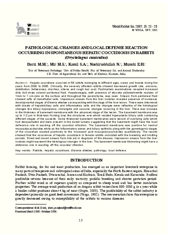
Pathological changes and local defense reaction occurring in spontaneous hepatic coccidiosis in rabbits (Oryctolagus cuniculus)
Mostrar el registro completo del ítem
Darzi, M.; Mir, M.; Kamil, S.; Nashiruddullah, N.; Munshi, Z. (2007). Pathological changes and local defense reaction occurring in spontaneous hepatic coccidiosis in rabbits (Oryctolagus cuniculus). World Rabbit Science. 15(1):23-28. https://doi.org/10.4995/wrs.2007.608
Por favor, use este identificador para citar o enlazar este ítem: http://hdl.handle.net/10251/8212
Ficheros en el ítem
Metadatos del ítem
| Título: | Pathological changes and local defense reaction occurring in spontaneous hepatic coccidiosis in rabbits (Oryctolagus cuniculus) | |
| Autor: | Darzi, M.M. Mir, M.S. Kamil, S.A. Nashiruddullah, N. Munshi, Z.H. | |
| Fecha difusión: |
|
|
| Resumen: |
[EN] Hepatic coccidiosis occurred in 56 rabbits belonging to different ages, sexes and breeds during the years from 2002 to 2005. Clinically, the severely affected rabbits showed decreased growth rate, anorexia, debilitation, ...[+]
|
|
| Palabras clave: |
|
|
| Derechos de uso: | Reserva de todos los derechos | |
| Fuente: |
|
|
| DOI: |
|
|
| Editorial: |
|
|
| Versión del editor: | https://doi.org/10.4995/wrs.2007.608 | |
| Tipo: |
|


